Comprehensive Eye Health Guide: Understanding Symptoms, Causes, and Preventative Care
Eye health refers to the overall condition of the visual system, including structures such as the cornea, lens, retina, and optic nerve, and it determines how well we see, interact with the world, and maintain quality of life. This guide explains common eye disease symptoms, biological causes, prevention strategies, digital eye strain solutions, emerging treatments, and practical vision correction options so readers can recognize warning signs and take action. ARY News is committed to informing and educating the public on health topics, and this article forms part of broader awareness coverage intended to help readers identify risks, find care, and follow reliable reporting. Many people experience treatable conditions like refractive error, dry eye, and early cataract yet delay care; clear guidance on symptoms, screening intervals, and lifestyle steps can reduce avoidable vision loss. The sections below walk through symptoms to watch for, major causes including diabetes and age-related changes, prevention tips and screening schedules, concrete relief for screen-related strain, recent technological advances in ophthalmology, the Pakistani context for eye health and youth-specific risks, and practical comparisons of vision correction choices. Read on to learn how to detect problems early, where to prioritize prevention, and how to weigh treatment and technology options for better vision.
What Are the Most Common Eye Disease Symptoms to Watch For?
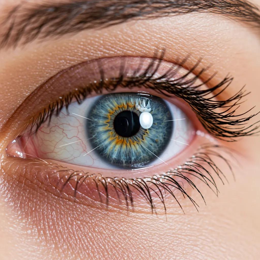
Common eye disease symptoms are warning signs that vision or ocular health is changing, and prompt evaluation can preserve sight. Blurred vision, sudden vision loss, new floaters or flashes, persistent eye pain, and increasing light sensitivity are among the top indicators that require attention. Because some sight-threatening conditions progress with little discomfort, regular screening is essential even when symptoms are absent. Understanding these universal signs helps readers decide when to seek urgent care and when to schedule routine assessment, which leads naturally into recognizing early, age-specific indicators and differentiating disease profiles below.
This list highlights the most frequent symptoms that indicate possible eye disease and when to act.
- Blurred or dimmed vision: Sudden or progressive reduction in clarity for near or distance tasks.
- Sudden vision loss: Rapid decrease in sight in one or both eyes — urgent evaluation needed.
- New floaters or flashes: Sudden appearance of floaters or light flashes may signal retinal detachment.
- Eye pain or severe redness: Can indicate infection, acute glaucoma, or inflammatory disease.
- Distorted central vision: Wavy or missing central areas suggest macular problems.
These symptoms form a practical checklist for readers to monitor and discuss with an eye professional, and recognizing them leads into specific early signs by age and condition.
Which Early Signs Indicate Vision Impairment?
Early signs of vision impairment vary by age but typically include subtle changes in visual tasks and behavior that progress over weeks to months. Children may sit too close to screens, tilt their head, or struggle with reading and classroom performance, while adults might begin to squint, need brighter light for tasks, or notice reduced night driving ability. Older adults often report gradual difficulty with fine detail, increased glare sensitivity, or trouble distinguishing faces, which can indicate age-related changes such as cataract or macular disease. If these patterns appear, scheduling an eye exam can identify refractive error or emerging pathology and enable timely intervention that preserves function and independence.
How Do Symptoms Differ Among Cataracts, Glaucoma, and Macular Degeneration?
Cataracts, glaucoma, and macular degeneration produce distinct symptom patterns that help clinicians differentiate them early in assessment and guide testing and management. Cataracts typically cause gradual clouding of vision, increased glare and halos around lights, and reduced color contrast, often worsening in low light. Glaucoma characteristically affects peripheral vision first and may be asymptomatic until advanced; increased intraocular pressure and optic nerve damage lead to tunnel vision if untreated. Macular degeneration primarily impairs central vision, producing distortions—straight lines appearing wavy—or central scotomas that hinder reading and facial recognition. Understanding these differences directs clinicians to specific diagnostics, such as slit-lamp exam for cataracts, visual field testing for glaucoma, and optical coherence tomography for macular disease.
What Are the Symptoms of Digital Eye Strain and How Can You Recognize Them?
Digital eye strain arises from prolonged screen exposure and presents with a cluster of symptoms that overlap with dry eye and refractive issues yet follow a characteristic time pattern. Typical complaints include eye fatigue, dryness, burning or gritty sensation, intermittent blurred vision, headaches, and neck or shoulder discomfort after extended device use.
Symptoms usually worsen toward the end of long screen sessions and improve with rest or after changing viewing habits, distinguishing strain from progressive ocular disease. Early recognition through symptom timing and simple self-checks enables practical interventions like scheduled breaks and ergonomic adjustments that reduce symptom burden and preserve visual comfort.
The extensive use of digital devices has led to a rise in digital eye strain, with a wide range of symptoms and prevalence reported.
Digital Eye Strain: A Comprehensive Review of Symptoms and Management
Digital eye strain (DES) is an entity encompassing visual and ocular symptoms arising due to the prolonged use of digital electronic devices. It is characterized by dry eyes, itching, foreign body sensation, watering, blurring of vision, and headache. Non-ocular symptoms associated with eye strain include stiff neck, general fatigue, headache, and backache. A variable prevalence ranging from 5 to 65% has been reported in the pre-COVID-19 era. With lockdown restrictions during the pandemic, outdoor activities were restricted for all age groups, and digital learning became the norm for almost 2 years. While the DES prevalence amongst children alone rose to 50–60%, the symptoms expanded to include recent onset esotropia and vergence abnormalities as part of the DES spectrum. New-onset myopia and increased progression of existing myopia became one of the most significant ocular health complications. Management options for DES include following correct ergonomics like reducing average daily screen time, frequent blinking, improving lighting, minimizing glare, taking regular breaks from the screen, changing focus to distance object intermittently, and following the 20-20-20 rule to reduce eye strain. Innovations in this field include high-resolution screens, inbuilt antireflective coating, matte-finished glass, edge-to-edge displays, and image smoothening graphic effects. Further explorations should focus on recommendations for digital screen optimization, novel spectacle lens technologies, and inbuilt filters to optimize visual comfort. A paradigm shift is required in our understanding of looking at DES from an etiological perspective, so that customized solutions can be explored accordingly. The aim of this review article is to understand the pathophysiology of varied manifestations, predisposing risk factors, varied management options, along with changing patterns of DES prevalence post COVID-19.
Digital eye strain-a comprehensive review, K Kaur, 2022
What Causes Vision Impairment and Common Eye Conditions?
Vision impairment stems from a mix of age-related degeneration, systemic disease, genetic predisposition, and environmental or behavioral factors, each contributing through distinct mechanisms. Age causes structural changes such as lens opacification and macular cell loss; systemic illnesses like diabetes damage retinal vessels; genetic and anatomical factors raise glaucoma risk through optic nerve vulnerability; and lifestyle elements—UV exposure, smoking, prolonged screens—accelerate disease or exacerbate symptoms. Identifying the causal pathway informs prevention and treatment choices, and the table below compares three leading causes with their primary mechanisms and risk contributors for clearer clinical perspective.
This comparison shows how age, systemic disease, and ocular physiology interact to produce distinct clinical problems, guiding targeted screening and management choices that follow.
What Are the Main Risk Factors for Cataracts and Glaucoma?
Cataracts and glaucoma share some risk features like increasing age, but they also have specific modifiable and non-modifiable risk factors that influence prevention strategies. For cataracts, aging is the dominant driver, while UV exposure, cigarette smoking, and prior ocular trauma or inflammation accelerate lens clouding; controlling environmental exposure and smoking cessation help reduce risk. Glaucoma risk includes elevated intraocular pressure, family history indicating genetic predisposition, African or Asian ancestry in some subtypes, and vascular comorbidities; regular eye pressure checks and optic nerve assessment detect disease before irreversible loss. Distinguishing modifiable factors from inherited risk supports personalized prevention plans centered on screening and lifestyle modification.
How Does Diabetes Lead to Diabetic Retinopathy?
Diabetic retinopathy results from chronic hyperglycemia damaging retinal blood vessels, causing microaneurysms, leakage, ischemia, and, in advanced stages, proliferative neovascularization that threatens vision. High blood sugar alters capillary walls and promotes inflammation and oxidative stress, leading to fluid accumulation in the macula and progressive retinal dysfunction; without control, bleeding or retinal detachment can occur. Staging ranges from background retinopathy to clinically significant macular edema and proliferative disease, and frequent retinal screening for people with diabetes is essential because early stages are often asymptomatic. Tight glycemic, blood pressure, and lipid control reduces incidence and progression, demonstrating how systemic disease management directly influences ocular outcomes.
The increasing global prevalence of diabetes necessitates robust screening for diabetic retinopathy, with artificial intelligence emerging as a key tool in this effort.
AI for Diabetic Retinopathy Screening: A Comprehensive Review
Diabetes is a global eye health issue. Given the rising in diabetes prevalence and ageing population, this poses significant challenge to perform diabetic retinopathy (DR) screening for these patients. Artificial intelligence (AI) using machine learning and deep learning have been adopted by various groups to develop automated DR detection algorithms. This article aims to describe the state-of-art AI DR screening technologies that have been described in the literature, some of which are already commercially available. All these technologies were designed using different training datasets and technical methodologies. Although many groups have published robust diagnostic performance of the AI algorithms for DR screening, future research is required to address several challenges, for examples medicolegal implications, ethics, and clinical deployment model in order to expedite the translation of these novel technologies into the healthcare setting.
Artificial intelligence for diabetic retinopathy screening: a review, A Grzybowski, 2020
Artificial intelligence, particularly deep learning, is proving instrumental in enhancing the accuracy and efficiency of diabetic retinopathy screening.
Diabetic Retinopathy Screening: The Role of Artificial Intelligence
Diabetic retinopathy is a frequent complication in diabetes and a leading cause of visual impairment. Regular eye screening is imperative to detect sight-threatening stages of diabetic retinopathy such as proliferative diabetic retinopathy and diabetic macular oedema in order to treat these before irreversible visual loss occurs. Screening is cost-effective and has been implemented in various countries in Europe and elsewhere. Along with optimised diabetes care, this has substantially reduced the risk of visual loss. Nevertheless, the growing number of patients with diabetes poses an increasing burden on healthcare systems and automated solutions are needed to alleviate the task of screening and improve diagnostic accuracy. Deep learning by convolutional neural networks is an optimised branch of artificial intelligence that is particularly well suited to automated image analysis. Pivotal studies have demonstrated high sensitivity and specificity for classifying advanced stages of diabetic retinopathy and identifying diabetic macular oedema in optical coherence tomography scans. Based on this, different algorithms have obtained regulatory approval for clinical use and have recently been implemented to some extent in a few countries.
Diabetic retinopathy screening in the emerging era of artificial intelligence, J Grauslund, 2022
What Environmental and Lifestyle Factors Contribute to Digital Eye Strain?
Digital eye strain arises from a constellation of environmental and behavioral contributors that converge during prolonged device use and poor workstation setup. Key factors include extended uninterrupted screen time, reduced blink rate causing tear film instability, poor posture and ergonomics, high screen brightness or contrast, and blue-enriched lighting during evening use that can disrupt sleep. Youth behaviors such as heavy social-media browsing, late-night device use, and near work at close distances intensify strain risk among 16–32-year-olds. Addressing these contributors with scheduling, ergonomic changes, and lighting adjustments provides practical relief and reduces cumulative ocular stress over time.
How Can You Prevent Eye Diseases and Maintain Good Eye Health?

Prevention of eye disease combines nutrition, routine eye exams, lifestyle changes, and protective behaviors; these steps reduce risk and preserve vision across the lifespan. A balanced diet rich in macular pigments and omega-3 fatty acids supports retinal health, while regular screening catches asymptomatic disease early. Lifestyle changes like smoking cessation, UV protection with sunglasses, managing chronic conditions such as diabetes, and moderating screen time all decrease long-term risk. The list below summarizes practical preventive actions readers can adopt immediately to protect vision and maintain ocular comfort.
- Eat nutrient-rich foods: Include leafy greens, fatty fish, and vitamin A sources to support retinal and corneal health.
- Schedule regular eye exams: Follow age- and risk-based screening intervals to detect asymptomatic conditions early.
- Protect from UV exposure: Wear sunglasses and hats outdoors to reduce cataract and ocular surface risk.
- Manage systemic disease: Control blood sugar, blood pressure, and cholesterol to lower retinopathy and vascular risk.
- Quit smoking: Smoking accelerates cataract and macular degeneration; cessation improves long-term outcomes.
- Adopt healthy screen habits: Use breaks, proper lighting, and ergonomic setups to prevent strain and dryness.
These practical measures form a foundation for lifelong eye care; the following subsections explain nutrient roles, exam frequency, and lifestyle changes that further reduce vision loss risk.
ARY News features health explainers and occasional interviews that highlight prevention campaigns and local screening initiatives; readers can follow ARY News coverage for updates on community programs and awareness drives. Positioning journalism as a public information resource complements clinical guidance and supports ongoing public education about screening and prevention.
What Are the Best Foods and Nutrients for Eye Health?
Certain nutrients support retinal structure, tear film stability, and overall ocular resilience, and including them in a balanced diet contributes to preventive eye care. Lutein and zeaxanthin, found in leafy greens, accumulate in the macula and help filter high-energy light, reducing age-related macular degeneration risk; omega-3 fatty acids from oily fish support retinal cell membranes and may ease dry eye symptoms. Vitamin A is essential for photoreceptor function and night vision, while antioxidants like vitamin C and E protect against oxidative stress linked to cataract and macular disease. Emphasizing whole-food sources and consulting clinicians before starting supplements helps readers apply nutritional strategies safely and effectively.
How Often Should You Have Regular Eye Exams for Early Detection?
Screening intervals vary by age and risk: children need early baseline exams and school screening participation, adults should have periodic evaluations that increase with age, and people with diabetes or other risk factors require annual or more frequent retinal exams. A typical framework includes an early childhood vision check, baseline adult exam in the 20s–30s, more frequent assessments after age 40–50 when presbyopia and age-related disease become more common, and annual diabetic retinal screening for people with diabetes. Local access issues and resource availability affect implementation, so readers should prioritize timely exams when symptoms or risk factors are present. Regular exams enable early detection of asymptomatic conditions like glaucoma and diabetic retinopathy, which preserves vision outcomes when treated early.
What Lifestyle Changes Help Reduce the Risk of Vision Loss?
Sustained lifestyle changes produce measurable benefits for eye health by reducing disease-promoting exposures and improving systemic control. Stopping smoking lowers the risk of cataract and macular degeneration, while ultraviolet protection through sunglasses limits lens and ocular surface damage. Effective management of chronic diseases—particularly blood glucose and hypertension—reduces progression of diabetic retinopathy and vascular ocular events. Finally, workplace ergonomics, sleep hygiene, and controlled screen time reduce chronic strain and dryness, creating a comprehensive prevention approach that preserves vision and daily function over time.
What Are Effective Solutions for Digital Eye Strain?
Digital eye strain responds well to a combination of behavioral routines, ergonomic adjustments, and targeted ocular therapies, so practical steps often bring rapid relief. The cornerstone routine is structured breaks with ocular exercises, optimizing device settings, and using lubricating drops when needed; these approaches address the underlying mechanisms of reduced blink rate, accommodative fatigue, and glare.
Use this simple three-step routine to relieve and prevent digital eye strain:
- 20-20-20 rule: Every 20 minutes, look at an object 20 feet away for 20 seconds to relax accommodation.
- Adjust screen settings and ergonomics: Reduce glare, match brightness to ambient light, and place screens at arm’s length and slightly below eye level.
- Maintain tear film and blinking: Perform blink exercises and use preservative-free artificial tears if dryness persists.
When these steps are applied consistently, many users see symptom reduction within days to weeks, while persistent or severe symptoms warrant professional evaluation to exclude other ocular conditions.
Which Habits and Exercises Relieve Digital Eye Strain Symptoms?
Daily habits like deliberate blinking, scheduled focusing shifts, and short palming or eye-rolling exercises reduce muscular fatigue and improve tear distribution during extended device use. Implementing micro-breaks—brief pauses every 20–30 minutes—to stand and perform eye-focus changes relieves accommodative strain, while posture corrections and chair adjustments reduce musculoskeletal contributions to headaches and discomfort. These exercises are low-risk, easy to integrate into work routines, and can be combined with the 20-20-20 rule for compounding benefit. If exercises do not improve symptoms, assessment for dry eye, uncorrected refractive error, or binocular vision dysfunction is appropriate to guide targeted therapy.
How Do Screen Settings and Breaks Improve Eye Comfort?
Adjusting screen brightness, contrast, and color temperature to match ambient lighting reduces glare and visual stress, while increasing text size and using high-contrast themes ease reading for prolonged periods. Warm color temperature settings during evening hours reduce circadian disruption and subjective visual discomfort, and software timers that prompt breaks help maintain consistent habits. Proper monitor distance—roughly an arm’s length—and height slightly below eye level minimizes upward gaze that exacerbates dryness. Together, these technical and behavioral adjustments decrease strain and support sustained productivity without compromising ocular comfort.
What Role Do Eye Drops and Artificial Tears Play in Managing Dry Eyes?
Artificial tears and lubricating eye drops restore tear film stability and relieve symptoms for many with mild to moderate dry eye, providing symptomatic relief that complements behavioral and ergonomic changes. Preservative-free formulations are preferable for frequent use to avoid cumulative preservative toxicity, while thicker gels or ointments help overnight symptom control for more severe cases. Over-the-counter drops address surface lubrication but do not treat underlying causes like Meibomian gland dysfunction, ocular allergy, or inflammatory disease; persistent symptoms should prompt clinician evaluation for prescription therapies or procedural options. Judicious use of lubricants as part of a broader management plan maintains comfort and supports adherence to digital-screen habits.
What Are the Latest Advances in Eye Health Technology and Treatments?
Recent innovations in ophthalmology improve early detection, monitoring, and procedural precision, expanding options for patients and clinicians alike. Artificial intelligence assists image interpretation and screening triage, smart contact lenses pursue continuous physiologic monitoring, and laser and robotic-assisted surgeries enhance surgical accuracy and recovery. These technologies serve different roles—from population-level screening to individual disease management—and each carries distinct maturity, benefit, and validation profiles that influence adoption. The table below summarizes selected advances, their primary attributes, and current clinical status to clarify how they contribute to improved outcomes.
This overview demonstrates how technology augments screening and treatment workflows, enabling earlier intervention and individualized care while requiring ongoing validation and clinical oversight.
How Is AI Enhancing Early Diagnosis of Eye Diseases?
AI enhances early diagnosis by analyzing retinal images and OCT scans to detect patterns that predict diabetic retinopathy, macular degeneration, and other retinal changes with high sensitivity in screening contexts. By automating initial interpretation, validated AI tools can triage large patient cohorts efficiently, enabling scarce specialist resources to focus on confirmed or complex cases. However, AI algorithms require clinical oversight, diverse training datasets, and regulatory validation to ensure accuracy across populations and imaging modalities. When integrated thoughtfully, AI supports earlier detection and referral pathways, particularly in resource-limited or outreach screening programs.
What Are Smart Contact Lenses and Their Benefits for Monitoring Eye Health?
Smart contact lenses embed sensors to track physiologic metrics such as intraocular pressure trends or tear-film biomarkers, offering the promise of continuous, unobtrusive monitoring for chronic ocular conditions. This monitoring could enable earlier detection of disease fluctuations and improved therapy titration for conditions like glaucoma, where pressure variation matters. Most smart lens applications remain in research or early clinical trials, and widespread clinical use depends on further validation, user comfort, data security, and regulatory clearance. As prototypes mature, smart lenses may complement clinic-based testing with real-world longitudinal data that informs personalized care.
How Are Laser and Robotic-Assisted Surgeries Improving Vision Correction?
Advances in femtosecond lasers and robotic assistance increase surgical precision for corneal and intraocular procedures, enabling more reproducible tissue sculpting and customized corrections that match individual anatomy. These technologies reduce mechanical variability, which can translate into improved visual outcomes, more predictable refractive results, and potentially faster recovery for selected patients. Suitability depends on ocular anatomy, coexisting disease, and patient expectations, and clinicians must balance benefits against procedural risks and candidacy criteria. As techniques evolve, careful outcome tracking and patient selection remain essential to realize consistent improvements in safety and vision restoration.
How Does Eye Health Affect the Pakistani Population and Youth?
Eye health in Pakistan reflects a mix of high-prevalence treatable conditions, access barriers, and emerging youth-specific challenges linked to digital habits and rising diabetes rates. Cataracts and uncorrected refractive error remain leading contributors to visual impairment, while diabetic retinopathy is increasing alongside national diabetes prevalence. Young people aged 16–32 face growing burdens of digital eye strain, myopia progression, and lifestyle-related risk behaviors that demand targeted awareness and school- or workplace-focused interventions. Reporting and public education can bridge information gaps, promote screening campaigns, and spotlight solutions that improve access and outcomes nationwide.
This local perspective underscores the importance of surveillance, outreach, and targeted prevention to reduce avoidable vision loss across demographic groups in Pakistan.
What Are the Most Common Eye Conditions in Pakistan?
Clinically, cataracts, uncorrected refractive errors, and diabetic retinopathy contribute substantially to vision impairment in Pakistan, reflecting global patterns modified by local access and disease trends. Cataract surgery backlog and unmet refractive correction needs disproportionately affect rural and low-income populations, while rising rates of diabetes drive increased retinal disease among working-age adults. Limited population-based data in some areas creates gaps in precise prevalence estimates, which reinforces the need for strengthened surveillance and program evaluation. Identifying these priorities enables targeted public health responses such as screening camps, school vision checks, and integration of eye care into primary health services.
What Challenges Exist in Accessing Eye Care Services Locally?
Access to eye care in Pakistan is constrained by geographic concentration of specialists in urban centers, financial barriers for surgery and corrective devices, and variability in public health outreach capacity. Rural populations often face long travel distances and limited local diagnostic services, while cost and awareness limit uptake of cataract surgery and refractive correction. Telemedicine, mobile eye camps, and public–private partnerships are promising strategies to extend reach, but sustainable funding and coordination are necessary to scale these approaches. Addressing workforce distribution, subsidized service models, and community education can reduce inequities and improve early detection and treatment uptake.
Which Public Awareness Campaigns Promote Eye Health in Pakistan?
Public awareness efforts in Pakistan include school screening initiatives, cataract surgical camps, and NGO-led education drives that emphasize early detection, spectacle provision, and diabetes-related eye care; these campaigns increase service uptake when combined with accessible follow-up care. Media coverage and community outreach that explain screening schedules, promote protective behaviors, and demystify surgical options help reduce stigma and delay in seeking care. ARY News has a role in highlighting these initiatives through reporting and video explainers that raise awareness and encourage participation in local programs, supporting sustained public engagement and accountability for eye health services.
What Should You Know About Vision Correction Options for Common Eye Conditions?
Vision correction spans non-invasive options like eyeglasses and contact lenses through to surgical interventions such as LASIK and cataract extraction, each with characteristic outcomes, recovery expectations, and suitability criteria. Eyeglasses offer immediate, reversible correction with minimal risk, contact lenses provide functional freedom with hygiene considerations, LASIK reshapes the cornea for refractive errors with rapid visual recovery for suitable candidates, and cataract surgery replaces the cloudy lens with an artificial intraocular lens to restore clarity. Comparing these options by procedure type, recovery, and typical outcomes helps readers set realistic expectations and choose a pathway aligned with their needs, which the table below summarizes.
This comparison aids decision-making by aligning condition severity, patient preferences, and expected timelines for recovery and visual function.
How Do Eyeglasses and Contact Lenses Correct Myopia and Astigmatism?
Eyeglasses and contact lenses correct refractive errors by altering the focal point of incoming light so it lands precisely on the retina, restoring clear vision for myopia, hyperopia, and astigmatism. Toric contact lenses and specialized spectacle lenses address astigmatism by compensating for irregular corneal curvature, while single-vision and multifocal options manage distance and near needs. Proper professional refraction and fitting ensure optimal visual acuity and comfort, and adherence to contact lens hygiene reduces infection risk. For progressive myopia in youth, evidence-based management strategies may modify progression and should be discussed with an eye care professional.
What Is LASIK Surgery and What Should Patients Expect?
LASIK is a laser-based refractive procedure that reshapes the corneal stroma to correct myopia, hyperopia, and some astigmatism, providing rapid visual improvement for appropriately screened candidates. Preoperative evaluation assesses corneal thickness, topography, and ocular health to determine candidacy and predict outcomes; surgery itself typically involves minimal discomfort and quick visual recovery over days to weeks. Risks include dry eye, visual disturbances such as halos or glare, and rare complications affecting corneal integrity; informed consent and realistic expectations are essential. Postoperative follow-up ensures healing and identifies any need for enhancement or management of side effects.
When Is Cataract Surgery Recommended and What Is the Recovery Process?
Cataract surgery is recommended when lens clouding significantly impairs function or quality of life, rather than being based solely on age; functional impairment in daily activities guides timing. Modern phacoemulsification surgery with intraocular lens implantation usually restores clarity and often reduces dependence on glasses, with patients experiencing progressive visual improvement over days to weeks. Postoperative care includes short-term topical medications, activity modifications, and scheduled follow-up visits to monitor healing and refractive outcome. Choosing IOL type—monofocal, multifocal, or toric—depends on individual visual goals and ocular health, with counseling by the surgeon guiding appropriate selection.
ARY News continues to report on local eye health campaigns, screening drives, and policy developments, serving as an information hub that raises awareness and encourages readers to follow ongoing coverage of vision-related public health initiatives.
Conclusion
Maintaining optimal eye health is crucial for preserving vision and enhancing quality of life, especially in the face of common conditions like cataracts and digital eye strain. By understanding symptoms, causes, and effective prevention strategies, individuals can take proactive steps to safeguard their eyesight. Regular eye exams, a balanced diet, and lifestyle modifications play a vital role in reducing the risk of vision impairment. Stay informed and explore our resources to prioritize your eye health today.